Home » Without Label » Bone Cross Section Real - Bone Cross Section High Res Stock Images Shutterstock / We recognized bone cross section by filtering pixel levels of ct image
Bone Cross Section Real - Bone Cross Section High Res Stock Images Shutterstock / We recognized bone cross section by filtering pixel levels of ct image
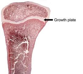
Bone Cross Section Real - Bone Cross Section High Res Stock Images Shutterstock / We recognized bone cross section by filtering pixel levels of ct image. The outlined area is a cross section of an osteon of compact bone. The previous image was correct, with one between the diaphysis and the head of the femur (which is an ossification center) and the other between the greater trochanter and the diaphysis. It consists of two layers; Translucence is an ivory characteristic that can be helpful in differentiating it from bone, as bone is an opaque substance. To the left is muscle tissue, and to the right is bone marrow.
To the left is muscle tissue, and to the right is bone marrow. An outer 'fibrous layer' containing mainly fibroblasts, and an inner 'cambium layer' containing progenitor cells. Illustration of the respiratory system: The only section of the proximal end of the femur that articulates is the head. It can be found under the periosteum and in the diaphyses of long bones, where it provides support and protection.
Bone Cross Section For Radius Digital Science On Behance from mir-s3-cdn-cf.behance.net The choice of ct versus mr depends on the structures and the disease processes that require assessment, delineation, and characterization. A central tube called a haversian canal typically runs in the same path as the length of the bone, and contains blood vessels, nerves, and. Elephant ivory always has schreger lines, a cross hatch pattern, when seen in cross section. 100x first focus in the compact decalcified bone (cb) on the left part of the image, you can see small dots, which are. The outside of a bone is covered in a thin layer of dense irregular connective tissue called the periosteum. Slice geometry will guess from your image title the bone it is working on. Cross section of a muscular artery showing the smooth muscle in the extensive tunica media, the endothelium and internal elastic membrane (lamina) which compose the. In three dimensions an osteon is cylindrical in shape.
Anyone dealing in ivory needs to know the laws regulating its sale, display and transportation.
Cross section of a muscular artery showing the smooth muscle in the extensive tunica media, the endothelium and internal elastic membrane (lamina) which compose the. The previous image was correct, with one between the diaphysis and the head of the femur (which is an ossification center) and the other between the greater trochanter and the diaphysis. If your bone of interest isn't listed, email me. 100x first focus in the compact decalcified bone (cb) on the left part of the image, you can see small dots, which are. Illustration of the respiratory system: Each ct frame image obtained in the previous step was resized by placing the cross section of bone in the centre of the frame. It consists of two layers; This slide contained a cross section of a very small bone, and you are looking at the entire thickness of the shaft of the bone. Slides have to be made this way because the matrix of bone is too hard to To the left is muscle tissue, and to the right is bone marrow. University of colorado museum of natural history object of the month october 2010 : Browse 3,292 human bone cross section stock photos and images available or start a new search to explore more stock photos and images. Looking at a bone in cross section, there are several distinct layered regions that make up a bone.
They are obtained by taking imaginary slices perpendicular to the main axis of organs, vessels, nerves, bones, soft tissue, or even the entire human body. In three dimensions an osteon is cylindrical in shape. On examining a section of any bone, it is seen to be composed of two kinds of tissue, one of which the marrow in the body of a long bone is supplied by one large artery (or sometimes more), which the canaliculi are exceedingly minute channels, crossing the lamellæ and connecting the lacunæ. In your example the area of cross section is quadrupled hence the bigger bar can take a four times larger force to extend by the. University of colorado museum of natural history object of the month october 2010 :
Science Update Bone Maturation Varies With Bone Size Suggest Nih Researchers Nichd Eunice Kennedy Shriver National Institute Of Child Health And Human Development from www.nichd.nih.gov A cross section of a compact bone shows concentric circles called lamellae. Each ct frame image obtained in the previous step was resized by placing the cross section of bone in the centre of the frame. Elephant ivory always has schreger lines, a cross hatch pattern, when seen in cross section. Browse 4,294 bone cross section stock photos and images available, or search for human bone cross section to find more great stock photos and pictures. An outer 'fibrous layer' containing mainly fibroblasts, and an inner 'cambium layer' containing progenitor cells. University of colorado museum of natural history object of the month october 2010 : Compact bone cross section courtesy: Compact bone is very different from the other tissues you have seen.
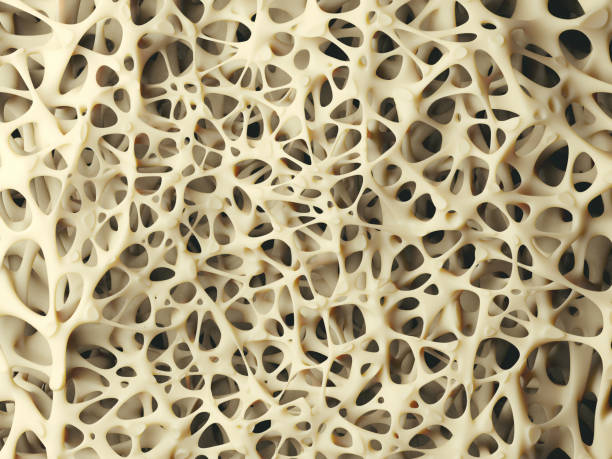
Compact bone is the denser, stronger of the two types of bone tissue ( (figure) ).
Concentric layers of bone cells (osteocytes) and bone matrix surround the central canal. A cross section of a compact bone shows concentric circles called lamellae. This is a short tutorial using blender 2.8 that shows how to create a bone cross section and using images to create the textures.hope you enjoy and please su. In the center of each osteon is the central canal, a space that houses blood vessels and nerves that supply bone. Foot bone anatomy x ray 12 photos of the foot bone anatomy x ray foot bone anatomy x ray, bone, foot bone anatomy x ray. Illustration of the respiratory system: Slice geometry will guess from your image title the bone it is working on. Smooth muscle and endothelium in a muscular artery wall, (magnification x100). This fits circles rather than spheres. The only section of the proximal end of the femur that articulates is the head. Looking at a bone in cross section, there are several distinct layered regions that make up a bone. On examining a section of any bone, it is seen to be composed of two kinds of tissue, one of which the marrow in the body of a long bone is supplied by one large artery (or sometimes more), which the canaliculi are exceedingly minute channels, crossing the lamellæ and connecting the lacunæ. Each ct frame image obtained in the previous step was resized by placing the cross section of bone in the centre of the frame.
This slide contained a cross section of a very small bone, and you are looking at the entire thickness of the shaft of the bone. This is a short tutorial using blender 2.8 that shows how to create a bone cross section and using images to create the textures.hope you enjoy and please su. Internal structure of a human long bone, with a magnified cross section of the interior. In the center of each osteon is the central canal, a space that houses blood vessels and nerves that supply bone. Compact bone is the denser, stronger of the two types of bone tissue ( (figure) ).
14 083 Bone Cross Section Stock Photos Pictures Royalty Free Images Istock from media.istockphoto.com This is a short tutorial using blender 2.8 that shows how to create a bone cross section and using images to create the textures.hope you enjoy and please su. Looking at a bone in cross section, there are several distinct layered regions that make up a bone. Cross section of a muscular artery showing the smooth muscle in the extensive tunica media, the endothelium and internal elastic membrane (lamina) which compose the. Foot bone anatomy x ray 12 photos of the foot bone anatomy x ray foot bone anatomy x ray, bone, foot bone anatomy x ray. It consists of two layers; Compact bone is the denser, stronger of the two types of bone tissue ( (figure) ). The only section of the proximal end of the femur that articulates is the head. An outer 'fibrous layer' containing mainly fibroblasts, and an inner 'cambium layer' containing progenitor cells.
It can be found under the periosteum and in the diaphyses of long bones, where it provides support and protection.
Browse 4,294 bone cross section stock photos and images available, or search for human bone cross section to find more great stock photos and pictures. Anyone dealing in ivory needs to know the laws regulating its sale, display and transportation. The upper (biting) surfaces of the tooth are at top, with the lower sections (bottom) embedded in the gums and jaw bone (not shown). Smooth muscle and endothelium in a muscular artery wall, (magnification x100). The previous image was correct, with one between the diaphysis and the head of the femur (which is an ossification center) and the other between the greater trochanter and the diaphysis. In the center of each osteon is the central canal, a space that houses blood vessels and nerves that supply bone. An outer 'fibrous layer' containing mainly fibroblasts, and an inner 'cambium layer' containing progenitor cells. After a fracture, woven bone forms initially and is gradually replaced by lamellar bone during a process known as bony substitution. They are obtained by taking imaginary slices perpendicular to the main axis of organs, vessels, nerves, bones, soft tissue, or even the entire human body. Foot bone anatomy x ray 12 photos of the foot bone anatomy x ray foot bone anatomy x ray, bone, foot bone anatomy x ray. In your example the area of cross section is quadrupled hence the bigger bar can take a four times larger force to extend by the. This fits circles rather than spheres. If it is wrong, correct it.
The choice of ct versus mr depends on the structures and the disease processes that require assessment, delineation, and characterization bone cross section. The upper (biting) surfaces of the tooth are at top, with the lower sections (bottom) embedded in the gums and jaw bone (not shown).